Rεςεαρch
42.81675,-73.926742
Enclosure transform for interest point detection from speckle imagery
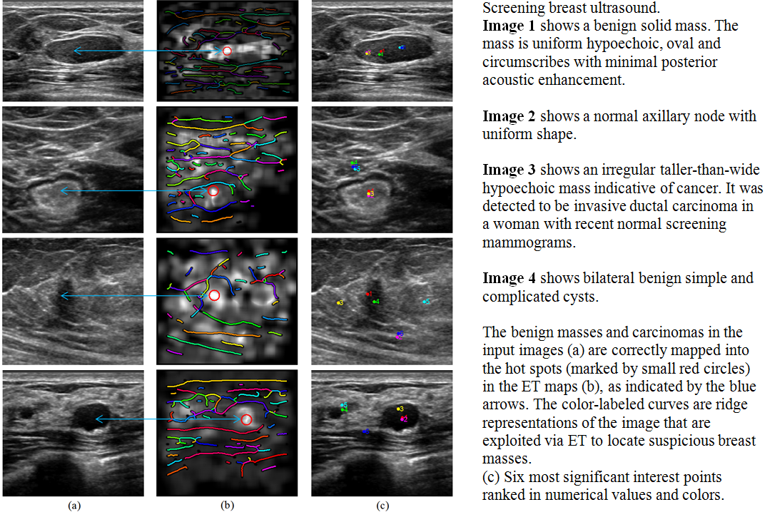
[paper]Automatic detection of points/objects of interest provides localizations of pathological appearance such as disease, cancer, in ammation, calcification, blood cells, and so on. The interest points can be used to reduce manual initialization effort and enable fully automatic object segmentation towards quantitative clinical analysis and more accurate pathological measurements.
We have devised a fast enclosure transform (ET) for localizing complex objects of interest from speckle imagery. Instead of relying on the sophisticated local properties of the object itself, ET makes use of regional features from a sparse image representation. Unrelated, broken ridge features surrounding an object are organized collaboratively, giving rise to the enclosureness of the object. We construct three enclosure likelihood measures, consisting of the enclosure force, potential energy, and encloser count. In the transform domain, the local maxima manifest the locations of interest objects, for which only the intrinsic dimension is known a priori. The discrete ET algorithm has a significant low computational cost and complexity.
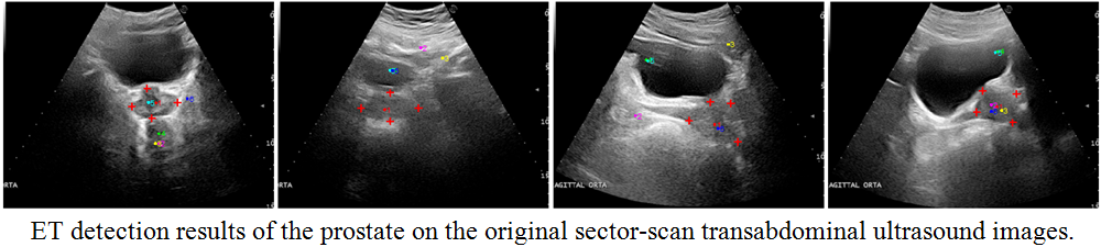
We apply ET to (1) detecting suspicious breast masses in screening breast ultrasound for women with dense breast tissue on mammography, and (2) automatic detection of the prostate locations from supra-pubic ultrasound images for verification of patient positioning in radiotherapy treatment of prostate cancer. ET yields superior results in terms of positive detection rate, accuracy and coverage. ET is a step forward towards robust automated image segmentation in speckle imagery which would otherwise demand human interventions.